PATIENT PRESENTATION
A 68 yo Caucasian male presented for evaluation and possible treatment.
CLINICAL DESCRIPTION
The patient was asymptomatic and the appearance of the mandibular left region was normal in color and contour. The mandibular left teeth tested normally to percussion, palpation, bite-stick, electric pulp test, thermal, periodontal and mobility. Patient denied history of trauma.
RADIOGRAPHIC DESCRIPTION OF LESION
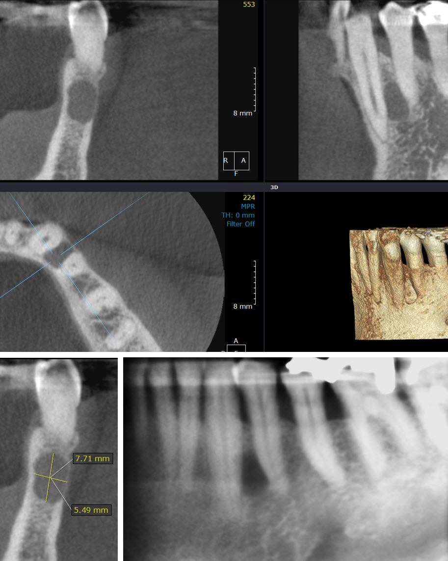
There is a 7 mm x 5 mm well defined, partially corticated teardrop shaped low density lesion in between the apical thirds of the mandibular left first bicuspid and cuspid, extending from lingual cortex to buccal cortex. The buccal cortex is uniformly and severely thinned, the lingual cortex is slightly thinned. There is no associated root resorption, however the lamina dura on the teeth of interest was effaced. The apices are not involved. Moderate periodontal bone loss on the visualized teeth.
(The listed structures are reviewed and evaluated for bilateral symmetry, configuration, cortical outline, medullary space, and patent sinuses/airways. Evaluation of the CBCT anatomical volume is intended as an overall review for pathology and abnormalities not directly associated with dental and periodontal conditions best imaged by conventional dental radiography. All viewed structures determined to have no significant findings are reported as no abnormalities detected.)
IMPRESSIONS AND RECOMMENDATIONS
- The low density lesion is suggestive of a lateral periodontal cyst between the mandibular left first bicuspid and cuspid. An excisional biopsy is indicated to confirm the diagnosis and rule out other pathosies.
- Moderate periodontal bone loss on the visualized teeth.
(The purpose of this image examination is to provide an evaluation of the regional anatomical volume not directly involved with the specific intent of the imaging examination. Evaluation is limited to the capability of CBCT imaging and any further assessment of dental related conditions is best performed by conventional dental radiography. This is a consultative report only and is not intended to be a definitive diagnosis or treatment plan.)
Consulting Radiologist:
Rujuta Katar, BDS, MDS, MS
Diplomate, American Board of Oral & Maxillofacial Radiology