Main Menu
CBCT Learning Center
for Endodontists
Upcoming Courses
October 23-24, 2026
Systematic Interpretation, Root Fractures, Management of Image Artifacts and Artificial Intelligence (Course 1)
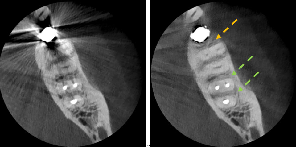
Newer metal‑artifact‑reduction (MAR) algorithms can be applied either during CBCT acquisition or as a post‑processing step to reduce streaking and distortion caused by highly attenuating materials. A, Axial CBCT slice reconstructed with a standard FDK algorithm. B, The identical dataset processed with a MAR algorithm demonstrates substantial artifact reduction along the mesial root of the mandibular first molar (yellow arrow) and the mesial and distal roots of the endodontically treated second molar (green arrows). Although MAR can improve visualization by suppressing beam‑hardening artifacts, it also subtracts voxel data within the affected region, which may obscure or alter diagnostically relevant structures.
Location: Washington, DC
Duration: 2 Days
More Course and Travel Information
Join us in Washington, DC for our 2‑day course on CBCT use in endodontics. This evidence‑based program applies to all limited field‑of‑view CBCT units and reflects the most current research and clinical guidance. Presented more than 80 times over the past 15 years, it is ideal for clinicians aiming to enhance diagnostic accuracy, refine interpretation skills, and elevate radiographic decision‑making.
Martin D. Levin, DMD, Barry Pass, DDS, PhD, and Thomas P. Winkler, MD
What You Will Learn
This comprehensive program blends scientific rigor with practical clinical guidance, covering essential topics including:
-
- Identifying Structural Fractures and Anomalies
- Vertical root fractures (VRF), specific bone loss patterns and radiographic features that are consistent or suggest a fracture
- Complex canal anatomy, mapping extra, missed or severely calcified canals
- Resorptive defects, diagnosis and management
- Systematic interpretation of CBCT scans, screening for incidental findingsArtifacts, metal artifact reduction (MAR), noise reduction and common interpretation errors
- Reporting on cross-sectional images, methods, and conventions
- Biologic effects of radiation dose relevant to endodontic assessments
- Comparative radiation doses for 2D and 3D
- Endodontic application of CBCT in endodontics
- Maxillary sinus anatomy, referring to otolaryngologist and endodontic considerations
- Radiologic anatomy of the CBCT image, normal and pathologic findings
- Principles of CBCT image production
- Workflow, the consent process, medico-legal considerations and branding
- Artificial intelligence, exploring the emerging role and responsible clinical use in endodontic assessments
- Identifying Structural Fractures and Anomalies
Who should attend
This course is ideal for endodontists, general dentists, residents, and specialists who use or plan to integrate CBCT in clinical practice and want a structured, evidence‑based approach to interpretation, reporting and responsible utilization.
Google Scholar Citations for Martin D. Levin, DMD
By The Numbers
82
Number of CBCT Courses presented by the CBCT Learning Center for Endodontists
More than 700
Number of Endodontists and Residents who have completed our program
5/5
Average rating by course attendees November 7-8, 2025
95%
Percentage of U.S. Endodontists with on-site CBCT units (Duong, 2023)
Case Studies
In The News
- Martin D. Levin, DMD Presented a Featured Lecture at APEC 2025 in Cairo
- The All India Institute of Medical Sciences, Center for Dental Education and Research will host Dr. Levin in New Dehli.
- Dr. Levin to Lecture at the 4th International Conference of the Bangladesh Endodontic Society, 2025, in Dhaka, Bangladesh
- Dr. Levin to Address the Marshall Baumgartner Endodontic Study Group in Portland, OR
- Dr. Martin D. Levin and Dr. Neil Starr Present “Preserving Your Dentition for Life” at the Cosmos Club’s Wellness Group
- AAE Publishes “A Different Kind of Case”
- Endodontic Society of the Philippines 37th Annual Meeting
- Ingle’s Endodontics, Ed 7 is Published
- Mid-Atlantic Archaeological Conference Paper Presented on Early Dentistry from a Native American Burial in the Southern Chesapeake Region, Virginia
- University of Pennsylvania, School of Dental Medicine Alumni Profile: Dr. Martin D. Levin, D’72, GD’74
EndoNet Consulting, LLC The Barlow Building 5454 Wisconsin Avenue, Suite 815 Chevy Chase, Maryland 20815-6901 Telephone: 301-654-6077 Fax: 301-654-0021 e-mail: info@endonet.com 15 CE Credits Awarded Teaching Method: Lecture Subject Code: 074
![]()
 Link to Google Books, Pathways of the Pulp, Chapter 2, Nair M, Levin M, Nair U. Radiographic Interpretation. In: Cohen’s Pathways of the Pulp by K. Hargreaves and L. Berman, ed. Mosby: St. Louis, Chp 2, 2020.
Link to AAE and AAOMR Joint Position Statement on CBCT in Endodontics 2025 Update