RADIOGRAPHIC FINDINGS:
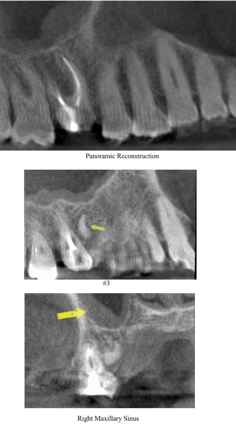
Partially visible right maxillary sinus: Mild opacification noted. #3: A curved, well-defined, high density lesion noted mesial to palatal root/palatal to MB root. It is ~ 6.5 mm in size.
(The listed structures are reviewed and evaluated for bilateral symmetry, configuration, cortical outline, medullary space, and patent sinuses/airways. Evaluation of the CBCT anatomical volume is intended as an overall review for pathology and abnormalities not directly associated with dental and periodontal conditions best imaged by conventional dental radiography. All viewed structures determined to have no significant findings are reported as no abnormalities detected.)
IMPRESSIONS AND RECOMMENDATIONS:
- Retained root possibly of #A/ idiopathic sclerosis, no associated pathology noted.
- Chronic Sinusitis of partially visible right maxillary sinus. No treatment is needed.
(The purpose of this image examination is to provide an evaluation of the regional anatomical volume not directly involved with the specific intent of the imaging examination. Evaluation is limited to the capability of CBCT imaging and any further assessment of dental related conditions is best performed by conventional dental radiography. This is a consultative report only and is not intended to be a definitive diagnosis or treatment plan.)
Consulting Radiologist: Marcel Noujeim, DDS, MS
Diplomate, American Board of Oral & Maxillofacial Radiology
