RADIOGRAPHIC FINDINGS:
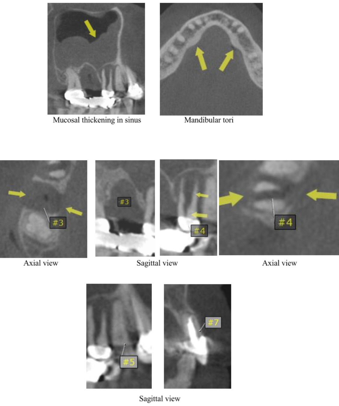
- Paranasal Sinuses: Polypoid shaped severe mucosal thickening of the right maxillary sinus.
- Nasal Cavities: No abnormalities detected
- Airway: No abnormalities detected
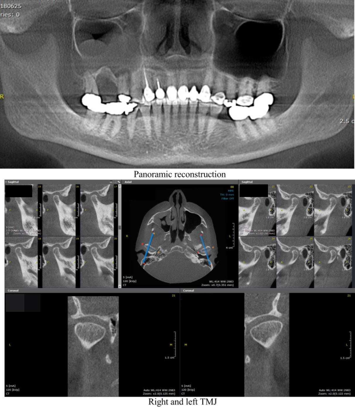
- Temporomandibular Joints: Both joints are well visualized and no abnormalities are detected
– Condyle: The condylar head is normal in size and shape with a smooth, continuous cortical outline and a normal underlying trabecular pattern with mild flattening noted in the superior aspect of the condylar head.
– Glenoid fossa and articular eminence: Glenoid fossa and eminence exhibit a normal bony contour and profile with no abnormalities detected. - Osseous Structures:
– Bilateral mandibular lingual tori.
– Moderate horizontal bone loss - Dental findings:
Tooth #3 area: Severe vertical bone loss extending from the mesial aspect of tooth #2 to the distal aspect of #4 with interruption of the buccal and palatal cortices, and elevation of the sinus floor without interruption.
Tooth #4: Well-defined oval shaped low density perradicular lesion extending from the mid third to the apex of the root, reaching the distal aspect of #5, with interruption of the buccal and palatal cortices, and causing elevation of the sinus floor without interruption.
– Vertical root fracture extending from the coronal third to the apex of the root.
(The listed structures are reviewed and evaluated for bilateral symmetry, configuration, cortical outline, medullary space, and patent sinuses/airways. Evaluation of the CBCT anatomical volume is intended as an overall review for pathology and abnormalities not directly associated with dental and periodontal conditions best imaged by conventional dental radiography. All viewed structures determined to have no significant findings are reported as no abnormalities detected.)
IMPRESSIONS AND RECOMMENDATIONS:
- Tooth #4: Poor prognosis, due to the vertical root fracture, extraction is needed.
- Tooth #5: The pulp canal is clear, patent and traceable along the length of the root.
- Tooth #7: Canal is obturated with adequate taper and density, and there is no radiographic evidence of periapical pathosis.
- Recent extraction socket in the area of #3 with no features suggestive of bone healing yet
(The purpose of this image examination is to provide an evaluation of the regional anatomical volume not directly involved with the specific intent of the imaging examination. Evaluation is limited to the capability of CBCT imaging and any further assessment of dental related conditions is best performed by conventional dental radiography. This is a consultative report only and is not intended to be a definitive diagnosis or treatment plan.)
Consulting Radiologist: Marcel Noujeim, DDS, MS
Diplomate, American Board of Oral & Maxillofacial Radiology